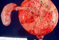
Choledochal cysts are congenital bile duct anomalies. These cystic dilatations of the biliary tree can involve the extrahepatic biliary radicles, the intrahepatic biliary radicles, or both. In 1723, Vater and Ezler published the anatomical description of a choledochal cyst. Douglas wrote the clinical report involving a 17-year-old girl presenting with jaundice, fever, intermittent abdominal pain, and an abdominal mass.[1] The patient died a month after an attempt at percutaneous drainage of the mass. (See image below.)
 Operative specimen of type I choledochal cyst. In 1959, Alonzo-Lej produced a systematic analysis of choledochal cysts, reporting on 96 cases. He devised a classification system, dividing choledochal cysts into 3 categories, and outlined therapeutic strategies. Todani has since refined this classification system to include 5 categories. This article reviews the incidence, pathophysiology, diagnosis, and management of choledochal cysts.
 Operative specimen of type I choledochal cyst. In 1959, Alonzo-Lej produced a systematic analysis of choledochal cysts, reporting on 96 cases. He devised a classification system, dividing choledochal cysts into 3 categories, and outlined therapeutic strategies. Todani has since refined this classification system to include 5 categories. This article reviews the incidence, pathophysiology, diagnosis, and management of choledochal cysts.
Recent research
In a retrospective analysis of 32 children and 47 adults with choledochal cysts, Shah et al investigated the differences between these 2 groups with regard to the presentation, management, and histopathology of, as well as the outcomes related to, these lesions.[2] The following were among the authors' findings:- A history of biliary surgery, pancreatitis, cholangitis, early postoperative complications, and late postoperative complications occurred, respectively, 5.1, 5.4, 6.4, 2.0, and 3.3 times more frequently in adults than they did in children.
- The classic triad of abdominal pain, jaundice, and a palpable right upper quadrant abdominal mass occurred 6.7 times more frequently in children than in adults.
- Fibrosis of the cyst wall was peculiar to children.
- Signs of inflammation and hyperplasia were primarily seen in adults.
- Long-term complications occurred in 29.7% of adults but in only 9.3% of children.
ليست هناك تعليقات:
إرسال تعليق