الجمعة، 3 فبراير 2012

Bile Duct Tumors

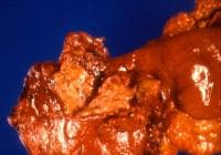
Tumors of the biliary tract (see image below) are uncommon but serious problems. The spectrum of lesions ranges from benign tumors, such as adenomas, to malignant lesions, such as adenocarcinomas. This discussion excludes tumors of the gallbladder, which are discussed separately.
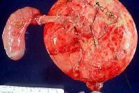
Distal common bile duct tumor excised by radical pDistal common bile duct tumor excised by radical pancreaticoduodenectomy. The tumor measured 1.2 cm in diameter.
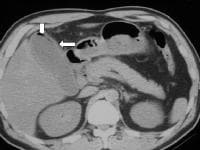
Most patients with bile duct tumors present with jaundice due to obstruction of the biliary tree by the tumor. Because the tumors are generally small, standard imaging studies, such as ultrasonography[1] and computed tomography (CT) scanning, may fail to show the lesion. These techniques may, however, provide a clue to the level of the obstruction and help exclude metastatic disease.
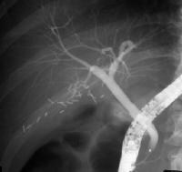
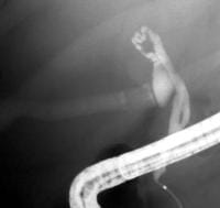
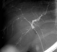
Cholangiography via a transhepatic or endoscopic approach is required to define the biliary anatomy and extent of the lesion. Magnetic resonance cholangiography is a noninvasive alternative available in an increasing number of centers.
The anticipated course of most cases of bile duct tumors includes recurrent biliary obstruction with infectious complications, local spread, and death in 6-12 months. Treatment depends on the site and extent of the lesion, and surgical resection improves survival and prognosis.

Benign Gastric Tumors

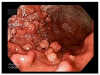
With the advent of modern techniques and the widespread use of gastric endoscopy, benign gastric wall lesions are now diagnosed more frequently and can be studied using the tissue obtained by biopsy or polypectomy. In the past, the diagnosis of gastric tumors was based on x-ray examination, but, in 1922, Schendler was the first to make an endoscopic-based diagnosis.[1] Note the image below.
Upper endoscopy showing multiple gastric polyps. Upper endoscopy showing multiple gastric polyps.

Acute Mesenteric Ischemia

Acute mesenteric ischemia (AMI) is a syndrome in which inadequate blood flow through the mesenteric circulation causes ischemia and eventual gangrene of the bowel wall. Broadly, AMI may be classified either as arterial or venous disease.
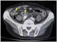
Arterial disease may be subdivided into nonocclusive mesenteric ischemia (NOMI; see the image below) and occlusive mesenteric arterial ischemia (OMAI). OMAI may be further subdivided into acute mesenteric arterial embolus (AMAE) and acute mesenteric arterial thrombosis (AMAT). Venous disease takes the form of mesenteric venous thrombosis (MVT). Thus, for practical purposes, AMI comprises 4 different primary clinical entities: NOMI, AMAE, AMAT, and MVT.
CT scan (with contrast) of nonocclusive mesentericCT scan (with contrast) of nonocclusive mesenteric ischemia with resulting bowel wall edema (arrows). The 4 types of AMI have somewhat different predisposing factors, clinical pictures, and prognoses. A secondary clinical entity of mesenteric ischemia occurs because of mechanical obstruction, such as internal hernia with strangulation, volvulus, intussusception, tumor compression, and aortic dissection. Occasionally, blunt trauma may cause isolated dissection of the superior mesenteric artery (SMA) and lead to intestinal infarction.
Because the 4 types of AMI share many similarities and a final common pathway (ie, bowel infarction and death, if not properly treated), they are discussed together.
In 1930, Cokkinis remarked, “Occlusion of the mesenteric vessels is apt to be regarded as one of those conditions of which the diagnosis is impossible, the prognosis hopeless, and the treatment almost useless.”[1] This quote indicates some of the extreme difficulties faced by physicians treating AMI. Symptoms are nonspecific initially, before evidence of peritonitis presents. Thus, diagnosis and treatment are often delayed until the disease is advanced.
Fortunately, since 1930, many advances have been made that allow earlier diagnosis and treatment. Whereas the prognosis remains grave for patients in whom the diagnosis is delayed until bowel infarction has already occurred, patients who receive the appropriate treatment in a timely manner are much more likely to recover.[2]

Abdominal Angina

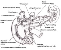
Abdominal angina is defined as the postprandial pain that occurs in individuals with sufficient mesenteric vascular occlusive disease such that blood flow cannot increase enough to meet visceral demands. The mechanism is believed to be similar to the angina pectoris that occurs in individuals with coronary artery disease or the intermittent claudication that accompanies peripheral vascular disease, as depicted in the image below.
The superior mesenteric artery and inferior mesentThe superior mesenteric artery and inferior mesenteric artery share collateral circulation near the splenic flexure of the colon. When dilated, this vessel is termed the meandering mesenteric artery. As seen on an angiogram, this is a sign of chronic mesenteric ischemia. The pancreaticoduodenal arcades are collateral patThe pancreaticoduodenal arcades are collateral pathways between the celiac artery and the superior mesenteric artery.
Although Schnitzler first described the clinical picture of postprandial clinical pain in 1901, description of the syndrome of postprandial abdominal angina generally is attributed to Baccelli or Goodman (1918). In 1936, Dunphy recognized that this syndrome was a precursor of fatal intestinal necrosis; however, not until 1957 did Mikkelsen propose surgical treatment of occlusive mesenteric vascular disease. Shaw and Maynard reported the first transarterial thromboendarterectomy of the superior mesenteric artery (SMA) in 1958, followed in rapid succession by Mikkelsen and Zarro in 1959. Numerous technical refinements followed.

Lower Gastrointestinal Bleeding

Lower gastrointestinal bleeding (LGIB) accounts for approximately 20-33% of episodes of gastrointestinal (GI) hemorrhage, with an annual incidence of about 20-27 cases per 100,000 population in Western countries. However, although LGIB is statistically less common than upper GI bleeding (UGIB), it has been suggested that LGIB is underreported because a higher percentage of affected patients do not seek medical attention.[1] Indeed, LGIB continues to be a frequent cause of hospital admission and is a factor in hospital morbidity and mortality LGIB is distinct from UGIB in epidemiology, management, and prognosis.
LGIB encompasses a wide spectrum of symptoms, ranging from trivial hematochezia to massive hemorrhage with shock. Acute LGIB is defined as bleeding that is of recent duration, originates beyond the ligament of Treitz, results in instability of vital signs, and is associated with signs of anemia with or without need for blood transfusion.
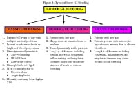
LGIB is classified under 3 groups according to the amount of bleeding, as shown in the image below. Massive hemorrhage is a life-threatening condition and requires transfusion of at least 5 U of blood.
Types of lower gastrointestinal (GI) bleeding. HR Types of lower gastrointestinal (GI) bleeding. HR = heart rate; SBP = systolic blood pressure.
Massive lower GI bleeding is defined as follows:
  • Passage of a large volume of red or maroon blood through the rectum
  • Hemodynamic instability and shock
  • Initial decrease in hematocrit (Hct) level of 6 g/dL or less
  • Transfusion of at least 2 U of packed red blood cells (RBCs)
  • Bleeding that continues for 3 days
  • Significant rebleeding in 1 week
LGIB has a mortality rate ranging from about 10-20%, with patients of advanced age (>60 y) and patients with comorbid conditions (eg, multiorgan system disease, transfusion requirements in excess of 5 units [U], need for operation, and recent stress, such as surgery, trauma, and sepsis) at greatest risk. LGIB is more likely in the elderly because of a higher incidence of diverticulosis and vascular disease in these groups. The incidence of LGIB is higher in men than in women.
Advances in diagnostic and therapeutic colonoscopy and in interventional angiography have resulted in a shift away from the need for surgical treatment (see the image below). Effective management with less invasive modalities has also reduced healthcare costs and, more importantly, patient morbidity and mortality.
Methods used to treat lower gastrointestinal bleedMethods used to treat lower gastrointestinal bleeding (LGIB).

Historical details

Understanding of the pathogenesis, diagnosis, and treatment of LGIB has drastically changed during the last 50 years. In the first half of the 20th century, large intestinal neoplasms were believed to be the most common cause of LGIB. In the 1950s, this condition was commonly attributed to diverticulosis; surgical treatment consisted of blind segmental bowel resections, with disappointing results. Patients who underwent these procedures suffered from a prohibitively high rebleeding rate (up to 75%), morbidity (up to 83%), and mortality (up to 60%).
In the last 4 decades, diagnostic methods for locating the precise bleeding point greatly improved. The flexible endoscope was developed in 1954. The full-length colonoscope was developed in 1965 in Japan. Also in 1965, Baum et al described selective mesenteric angiography, which permitted the identification of vascular abnormalities and the precise bleeding point.[2] The first anal colonoscopy was performed in 1969.
Experience with mesenteric angiography in the late 1960s and 1970s suggested that angiodysplasias and diverticulosis were the most common reasons for LGIB. Since its discovery, mesenteric angiography remains the criterion standard in precise localization of the bleeding.
Rosch et al described superselective visceral arteriography for infusion of vasoconstrictors in 1971 and superselective embolization of the mesenteric vessels as an alternative technique to treat massive LGIB in 1972.[3, 4] The most feared complication of embolization of the mesenteric vessels is ischemic colitis, which has limited its use for GI bleeding.
The initial experience with vasopressin infusion was reported in 1973-1974. Vasopressin causes vasoconstriction and arrests the bleeding in 36-100% of patients. The recurrence rate following completion of vasopressin infusion can be as high as 71%; therefore, vasopressin is used to temporize the acute event and to stabilize patients before surgery.
Endoscopic control of bleeding with thermal modalities or sclerosing agents has been in use since the 1980s. One of the advantages of upper (or lower) endoscopic evaluation is that it provides a means to administer therapy in patients with GI bleeding. Nuclear scintigraphy has been used since the early 1980s as a very sensitive diagnostic tool to evaluate bleeding from GI tract; this modality can detect hemorrhage at rates as low as 0.1 mL/min

Familial Adenomatous Polyposis

Familial adenomatous polyposis (FAP) is the most common adenomatous polyposis syndrome. It is an autosomal dominant inherited disorder characterized by the early onset of hundreds to thousands of adenomatous polyps throughout the colon. If left untreated, all patients with this syndrome develop colon cancer by age 35-40 years. In addition, an increased risk exists for the development of other malignancies. See the image below.
Colectomy specimen obtained from a patient with faColectomy specimen obtained from a patient with familial adenomatous polyposis. Note the presence of numerous synchronous adenomatous polyps lining the luminal surface. The genetic defect in FAP is a germline mutation in the adenomatous polyposis coli (APC) gene. Syndromes once thought to be distinct from FAP are now recognized to be, in reality, part of the phenotypic spectrum of FAP.
Syndromes with a germline mutation in the APC gene include FAP, Gardner syndrome, some families with Turcot syndrome, and attenuated adenomatous polyposis coli (AAPC). Gardner syndrome is characterized by colonic polyposis typical of FAP, along with osteomas (bony growth most commonly on the skull and the mandible), dental abnormalities, and soft tissue tumors. Turcot syndrome is characterized by colonic polyposis typical of FAP, along with central nervous system tumors (medulloblastoma). AAPC is characterized by fewer colonic polyps (average number of polyps, 30-35) as compared to classic FAP. The polyps also tend to develop at a later age (average age, 36 y), and they tend to involve the proximal colonic area.
In considering the spectrum of polyposis syndromes, patients with multiple adenomatous polyps most likely have FAP (or one of its variants), AAPC, or MYH-associated polyposis (MAP). If a patient with a suspected polyposis syndrome undergoes genetic testing and does not have an APC gene mutation, MYH gene testing should be performed to assess for MAP, as 10-20% of patients who do not have an APC gene mutation have biallelic MYH gene mutations.[1]
The phenotype of MAP is often indistinguishable from FAP or AAPC, with patients having usually 10-100 polyps but sometimes more than 100. The age of onset of MAP is usually in patients older than 45 years, and patients often present symptomatically, with colorectal carcinoma commonly found at the time of diagnosis. This is in part because there is usually no family history given the autosomal recessive inheritance pattern of MAP. Duodenal polyps can be found in up to one fifth of patients.[2] There is no increased risk of other types of cancers associated with this syndrome.

Schatzki Ring

Since the 1950s, several investigators have published reports of patients with dysphagia who had associated lower esophageal ringlike constrictions, but each investigator had a different opinion as to the cause and nature of these rings. In 1953, Ingelfinger and Kramer believed that these rings occurred as a result of a contraction by an overactive band of esophageal muscle[1] ; however, Schatzki and Gary believed that these rings were fixed and not contractile.[2] Some of this controversy may be related to the confusion of categorizing muscular and mucosal rings under the same entity, as concluded by Goyal et al.[3, 4, 5, 6]
Endoscopic appearance of the distal esophagus illuEndoscopic appearance of the distal esophagus illustrating a Schatzki ring.
Two rings have been identified in the distal esophagus. The muscular ring, or A ring, is a thickened symmetric band of muscle that forms the upper border of the esophageal vestibule and is located approximately 2 cm above the gastroesophageal junction. The A ring is rare; furthermore, it is even more rarely associated with dysphagia. On the other hand, the mucosal ring, or B ring, is quite common and is the subject of discussion in this article. The B ring is a diaphragmlike thin mucosal ring usually located at the squamocolumnar junction; it may be symptomatic or asymptomatic, depending on the luminal diameter.
The pathogenesis is not clear, and patients typically present with intermittent nonprogressive dysphagia for solids. Fortunately, most patients respond well to initial and repeat dilatation therapy. A small number of patients may have stubborn rings that require more aggressive endoscopic or surgical intervention.

Sengstaken-Blakemore Tube

Balloon tamponade of bleeding esophageal varices was described as early as the 1930s. A double-balloon tamponade system originally developed by Sengstaken and Blakemore in 1950 has undergone relatively few changes to the current day.[1, 2, 3] The 3 major components of a Sengstaken-Blakemore tube are a gastric balloon, an esophageal balloon, and a gastric suction port. The addition of an esophageal suction port to help prevent aspiration of esophageal contents resulted in what is called the Minnesota tube. Another nasogastric (NG) device with a single gastric balloon is most effective at terminating bleeding from gastric varices and is known as the Linton-Nachlas tube.[4] The advent of endoscopy has reduced the use of balloon tamponade, but the use of such devices can still be temporizing or lifesaving, despite their potential for serious complications.[5, 6, 7, 8]
See the images below.
Sengstaken-Blakemore tube. Image courtesy of RichaSengstaken-Blakemore tube. Image courtesy of Richard Treger, MD. Linton-Nachlas tube. Image courtesy of Richard TreLinton-Nachlas tube. Image courtesy of Richard Treger, MD.

Cytomegalovirus Colitis

Cytomegalovirus (CMV) is a member of the Herpesviridae family, along with herpes simplex viruses 1 and 2, Epstein-Barr virus, and varicella-zoster virus. It is a double-stranded DNA virus with a protein coat and lipoprotein envelope. Similar to other herpesviruses, CMV is icosahedral and replicates in the host's nucleus. Replication in the host cell typically manifests pathologically with large intranuclear inclusion bodies and smaller cytoplasmic inclusions, and is accompanied by presence of CMV viral particles in the plasma.
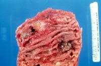
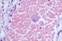
Gross specimen of bowel showing ulceration secondaGross specimen of bowel showing ulceration secondary to cytomegalovirus colitis. Giant cell with inclusion body characteristic of cGiant cell with inclusion body characteristic of cytomegalovirus colitis.
Between 50% and 80% of the world's population is seropositive for CMV. Initial CMV infection in the immunocompetent host typically is mild and goes undetected clinically. This is followed by a chronic latent state, during which the virus remains present within host cells, but viral proliferation is prevented by host cell-mediated immunity. Failure of immune containment may lead to reactivation with viral proliferation and severe systemic illness. Systemic CMV disease is characterized by fever, pancytopenia, and inflammatory changes in multiple organs including the liver and lungs, and in the retina. Colitis is a frequent manifestation of this acute systemic illness.
Patients are rendered susceptible to systemic CMV disease by treatment with immunosuppressive medications, or by illnesses that reduce cellular immunity, such as human immunodeficiency virus (HIV) infection. Acute systemic illness caused by CMV is particularly common following initial exposure in an immune compromised individual (in particular, in a CMV-negative transplant recipient who receives an organ from a CMV-positive donor).

Hirschsprung Disease

Hirschsprung disease is a developmental disorder of the enteric nervous system and is characterized by an absence of ganglion cells in the distal colon resulting in a functional obstruction.[1] See the image below.
Hirschsprung disease. Contrast enema demonstratingHirschsprung disease. Contrast enema demonstrating transition zone in the rectosigmoid region.
Although this condition was described by Ruysch in 1691 and popularized by Hirschsprung in 1886, the pathophysiology was not clearly determined until the middle of the 20th century, when Whitehouse and Kernohan described the aganglionosis of the distal intestine as the cause of obstruction in their series of patients.[2]
In 1949, Swenson described the first consistent definitive procedure for Hirschsprung disease, rectosigmoidectomy with coloanal anastomosis. Since then, other operations have been described, including the Duhamel and Soave techniques. More recently, advances in surgical technique, including minimally invasive procedures, and earlier diagnosis have resulted in decreased morbidity and mortality for patients with Hirschsprung disease.
Most cases of Hirschsprung disease are now diagnosed in the newborn period. Hirschsprung disease should be considered in any newborn who fails to pass meconium within 24-48 hours after birth. Although contrast enema is useful in establishing the diagnosis, full-thickness rectal biopsy remains the criterion standard. Once the diagnosis is confirmed, the basic treatment is to remove the poorly functioning aganglionic bowel and to create an anastomosis to the distal rectum with the healthy innervated bowel (with or without an initial diversion).

Pseudomembranous Colitis Surgery

Pseudomembranous colitis is an inflammatory disease of the colon (see image below). It has changed in the last 100 years from a fatal disease caused by a postoperative event to, in the era of antibiotics, a commonly occurring complication of antibiotic use that may lead to serious morbidity but that usually is treated easily.
Colonic pseudomembranes of pseudomembranous colitiColonic pseudomembranes of pseudomembranous colitis. Photographs courtesy of Eric M. Osgard, MD.
  • In the late 1800s, prior to the availability of antibiotics, Finney reported the first case of pseudomembranous colitis, calling it "diphtheritic colitis."[1]
  • Hall and O'Toole first described Clostridium difficile in 1935.[2]
  • C difficile was first implicated as a causative factor in pseudomembranous colitis in the 1970s

Toxic Megacolon

Toxic megacolon is the clinical term for an acute toxic colitis with dilatation of the colon. The dilatation can be either total or segmental. A more contemporary term for toxic megacolon is simply toxic colitis, because patients may develop toxicity without megacolon. For the purposes of this article, the term toxic megacolon (toxic colitis) is used, but either toxicity or megacolon can occur exclusively of each other.[1]
The hallmarks of toxic megacolon (toxic colitis), a potentially lethal condition, are nonobstructive colonic dilatation larger than 6 cm and signs of systemic toxicity. Toxic megacolon (toxic colitis)was recognized by Marshak and Lester in 1950.[2] Jalan et al described the diagnostic criteria.[3] The first criterion is radiographic evidence of colonic dilatation. The second criterion is any 3 of the following: fever (>101.5°F), tachycardia (>120 beats/min), leukocytosis (>10.5 103/µL), or anemia. The third criterion is any 1 of the following: dehydration, altered mental status, electrolyte abnormality, or hypotension.
Toxic megacolon (toxic colitis) was first thought to be a complication of ulcerative colitis. In fact, toxic megacolon (toxic colitis) may complicate any number of colitides, including inflammatory, ischemic, infectious, radiation, and pseudomembranous.[4, 5] See the image below.
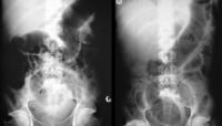
A 22-year-old man presented with abdominal pain, pA 22-year-old man presented with abdominal pain, passage of blood and mucus per rectum, abdominal distention, fever, and disorientation. Findings from sigmoidoscopy confirmed ulcerative colitis. Abdominal radiographs obtained 2 days apart show mucosal edema and worsening of the distention in the transverse colon. The patient's clinical condition deteriorated over the next 36 hours despite steroid and antibiotic therapy, and the patient had to undergo a total colectomy and ileostomy.
The incidence of toxic megacolon (toxic colitis) is expected to increase due to the rising prevalence of pseudomembranous colitis. Colonic dilatation may be present in other conditions, such as Hirschsprung disease, idiopathic megacolon/chronic constipation, and intestinal pseudo-obstruction (Ogilvie syndrome). However, these patients do not develop signs of systemic toxicity and, therefore, do not fall into the category of having toxic megacolon (toxic colitis). See the images below.
Gross pathology specimen from a case of pseudomembGross pathology specimen from a case of pseudomembranous colitis demonstrating characteristic yellowish plaques. Computed tomography scan from a patient with pseudComputed tomography scan from a patient with pseudomembranous colitis demonstrating the classic accordion sign.

Villous Adenoma

Adenomatous polyps are, by definition, neoplastic. Although benign, they are the direct precursors of adenocarcinomas and follow a predictable cancerous temporal course unless interrupted by treatment. They can be either pedunculated or sessile. Polyps are generally asymptomatic but may occasionally ulcerate and bleed; uncommonly, they may result in obstruction if very large. Adenomas are divided into 3 subtypes based on histologic criteria, as follows: (1) tubular, (2) tubulovillous, and (3) villous. According to World Health Organization (WHO) criteria, villous adenomas are composed of greater than 80% villous architecture. Tubular adenomas are encountered most frequently (80-86%). Tubulovillous adenomas are encountered less frequently (8-16%), and villous adenomas are encountered least frequently (5%).
See the images below.
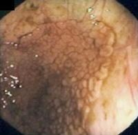
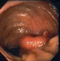
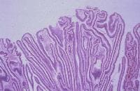
Endoscopic view of a sessile polyp, which histologEndoscopic view of a sessile polyp, which histology studies revealed to be a villous adenoma. Courtesy of H. Chaun, MD. Endoscopic view of a sessile polyp histologically Endoscopic view of a sessile polyp histologically determined to be a villous adenoma. Courtesy of R. Enns, MD. Histology of villous adenoma. Fingerlike projectioHistology of villous adenoma. Fingerlike projections stretching from the surface of a polyp downward with minimal branching. Courtesy of D. Owen, MD. Histology of villous adenoma. Low-grade dysplasia Histology of villous adenoma. Low-grade dysplasia with loss of mucin, prominent nucleoli, and hyperchromatic and elongated cells. Courtesy of D. Owen, MD.
Villous adenomas are associated more often with larger adenomas and more severe degrees of dysplasia. These adenomas occur more frequently in the rectum and rectosigmoid, although they may occur anywhere in the colon. They generally are sessile structures that appear as velvety or cauliflowerlike projections. Although rare, villous adenomas of the duodenum and the small bowel, particularly at the ampulla, can occur. Villous adenomas are of concern primarily because of the risk of malignant transformation (approximately 15-25% overall but higher once >2 cm).
The primary focus of this article is colonic villous adenomas. Where appropriate, certain aspects of small bowel villous adenomas are addressed

Bile Duct Strictures

Bile duct stricture (biliary stricture) is an uncommon but challenging clinical condition that requires a coordinated multidisciplinary approach involving gastroenterologists, radiologists, and surgical specialists. Unfortunately, most benign bile duct strictures (biliary strictures) are iatrogenic, resulting from operative trauma (see images below).[1] Bile duct strictures (biliary strictures) may be asymptomatic but, if ignored, can cause life-threatening complications, such as ascending cholangitis,[2, 3] liver abscess, and secondary biliary cirrhosis.
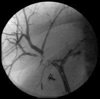
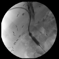
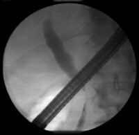
Focal intrahepatic benign bile duct stricture afteFocal intrahepatic benign bile duct stricture after cholecystectomy. Percutaneous transhepatic cholangiogram with balloPercutaneous transhepatic cholangiogram with balloon dilation of a postoperative bile duct stricture.
However, not all bile duct strictures (biliary strictures) are benign. Pancreatic cancer is the most common cause of malignant biliary strictures (see images below).[4, 5] Most of these patients die of complications of tumor invasion and metastasis rather than from the bile duct stricture (biliary stricture) per se. Nonetheless, both benign and malignant bile duct strictures can be associated with distressing symptoms and excessive morbidity.[6]
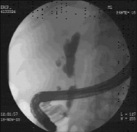
Endoscopic retrograde cholangiopancreatographic chEndoscopic retrograde cholangiopancreatographic cholangiogram demonstrating an isolated mid-hepatic duct stricture as a result of pancreatic cancer. Focal bile duct stricture as a result of pancreatiFocal bile duct stricture as a result of pancreatic cancer in the head of the pancreas.
For excellent patient education resources, visit eMedicine's Liver, Gallbladder, and Pancreas Center and Hepatitis Center. Also, see eMedicine's patient education articles Cirrhosis and Gallstones.

Bile Duct Tumors

Tumors of the biliary tract (see image below) are uncommon but serious problems. The spectrum of lesions ranges from benign tumors, such as adenomas, to malignant lesions, such as adenocarcinomas. This discussion excludes tumors of the gallbladder, which are discussed separately.
Distal common bile duct tumor excised by radical pDistal common bile duct tumor excised by radical pancreaticoduodenectomy. The tumor measured 1.2 cm in diameter.
Most patients with bile duct tumors present with jaundice due to obstruction of the biliary tree by the tumor. Because the tumors are generally small, standard imaging studies, such as ultrasonography[1] and computed tomography (CT) scanning, may fail to show the lesion. These techniques may, however, provide a clue to the level of the obstruction and help exclude metastatic disease.
Cholangiography via a transhepatic or endoscopic approach is required to define the biliary anatomy and extent of the lesion. Magnetic resonance cholangiography is a noninvasive alternative available in an increasing number of centers.
The anticipated course of most cases of bile duct tumors includes recurrent biliary obstruction with infectious complications, local spread, and death in 6-12 months. Treatment depends on the site and extent of the lesion, and surgical resection improves survival and prognosis.

Choledochal Cysts

Choledochal cysts are congenital bile duct anomalies. These cystic dilatations of the biliary tree can involve the extrahepatic biliary radicles, the intrahepatic biliary radicles, or both. In 1723, Vater and Ezler published the anatomical description of a choledochal cyst. Douglas wrote the clinical report involving a 17-year-old girl presenting with jaundice, fever, intermittent abdominal pain, and an abdominal mass.[1] The patient died a month after an attempt at percutaneous drainage of the mass. (See image below.)
Operative specimen of type I choledochal cyst. Operative specimen of type I choledochal cyst. In 1959, Alonzo-Lej produced a systematic analysis of choledochal cysts, reporting on 96 cases. He devised a classification system, dividing choledochal cysts into 3 categories, and outlined therapeutic strategies. Todani has since refined this classification system to include 5 categories. This article reviews the incidence, pathophysiology, diagnosis, and management of choledochal cysts.

Recent research

In a retrospective analysis of 32 children and 47 adults with choledochal cysts, Shah et al investigated the differences between these 2 groups with regard to the presentation, management, and histopathology of, as well as the outcomes related to, these lesions.[2] The following were among the authors' findings:
  • A history of biliary surgery, pancreatitis, cholangitis, early postoperative complications, and late postoperative complications occurred, respectively, 5.1, 5.4, 6.4, 2.0, and 3.3 times more frequently in adults than they did in children.
  • The classic triad of abdominal pain, jaundice, and a palpable right upper quadrant abdominal mass occurred 6.7 times more frequently in children than in adults.
  • Fibrosis of the cyst wall was peculiar to children.
  • Signs of inflammation and hyperplasia were primarily seen in adults.
  • Long-term complications occurred in 29.7% of adults but in only 9.3% of children.
The authors concluded that because of such differences, choledochal cysts in children should be considered separate entities from those in adults.

Cholelithiasis

Cholelithiasis is the medical term for gallstone disease. Gallstones are concretions that form in the biliary tract, usually in the gallbladder (see the image below).
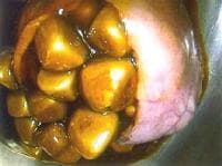
Cholelithiasis. A gallbladder filled with gallstonCholelithiasis. A gallbladder filled with gallstones (examined extracorporally after laparoscopic cholecystectomy [LC]).
Gallstones develop insidiously, and they may remain asymptomatic for decades. Migration of a a gallstone into the opening of the cystic duct may block the outflow of bile during gallbladder contraction. The resulting increase in gallbladder wall tension produces a characteristic type of pain (biliary colic). Cystic duct obstruction, if it persists for more than a few hours, may lead to acute gallbladder inflammation (acute cholecystitis).
Choledocholithiasis refers to the presence of one or more gallstones in the common bile duct. Usually, this occurs when a gallstone passes from the gallbladder into the common bile duct (see the image below).
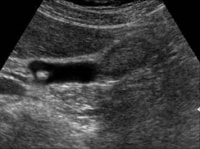
Common bile duct stone (choledocholithiasis). The Common bile duct stone (choledocholithiasis). The sensitivity of transabdominal ultrasonography for choledocholithiasis is approximately 75% in the presence of dilated ducts and 50% for nondilated ducts. Image courtesy of DT Schwartz.
A gallstone in the common bile duct may impact distally in the ampulla of Vater, the point where the common bile duct and pancreatic duct join before opening into the duodenum. Obstruction of bile flow by a stone at this critical point may lead to abdominal pain and jaundice. Stagnant bile above an obstructing bile duct stone often becomes infected, and bacteria can spread rapidly back up the ductal system into the liver to produce a life-threatening infection called ascending cholangitis. Obstruction of the pancreatic duct by a gallstone in the ampulla of Vater also can trigger activation of pancreatic digestive enzymes within the pancreas itself, leading to acute pancreatitis.[1, 2]
Chronically, gallstones in the gallbladder may cause progressive fibrosis and loss of function of the gallbladder, a condition known as chronic cholecystitis. Chronic cholecystitis predisposes to gallbladder cancer.
Ultrasonography is the initial diagnostic procedure of choice in most cases of suspected gallbladder or biliary tract disease (see Workup).
The treatment of gallstones depends upon the stage of disease. Asymptomatic gallstones may be managed expectantly. Once gallstones become symptomatic, definitive surgical intervention with excision of the gallbladder (cholecystectomy) is usually indicated. Cholecystectomy is among the most frequently performed abdominal surgical procedures (see Treatment). Complications of gallstone disease may require specialized management to relieve obstruction and infection.
Go to Pediatric Cholelithiasis for complete information on this topic.

Emphysematous Cholecystitis

Emphysematous cholecystitis, known less commonly as clostridial cholecystitis, is an acute infection of the gallbladder wall caused by gas-forming organisms (eg, Clostridium or Escherichia coli) that is generally considered a surgical emergency. An infrequent, insidious, and rapidly progressive form of acute cholecystitis, emphysematous cholecystitis is characterized by early gangrene, perforation of the gallbladder and high mortality. Although this condition develops in approximately 1% of all cases of acute cholecystitis, compared with typical acute cholecystitis, emphysematous cholecystitis is associated with much higher rates of gangrene and perforation of the gallbladder and significantly increased rates of mortality (15-25%).
An estimated 500,000 cholecystectomies are performed per year in the United States. Assuming all patients with emphysematous cholecystitis come to surgery, this would indicate that 5000 cholecystectomies are performed per year for emphysematous cholecystitis. Although the number of patients who are treated successfully without surgery is certainly small, the number of patients who die without surgery is unknown.
Usually, the diagnosis is made by the radiographic presence of air within the gallbladder wall or lumen. Elderly males, especially diabetics, are particularly susceptible to clostridial cholecystitis (emphysematous cholecystitis). The computed tomography (CT) scan below shows a patient with emphysematous cholecystitis.
Emphysematous cholecystitis in a 47-year-old man wEmphysematous cholecystitis in a 47-year-old man with diabetes who experienced abdominal pain. This computerized tomography scan shows gas within the wall of the gallbladder (horizontal arrow) as well as within the lumen of the gallbladder (vertical arrow). Radiograph courtesy of Helen Morehouse, MD.

Gallbladder Tumors

Gallbladder tumors are recognized with increasing frequency due to improvements in imaging techniques and increased utilization of these studies. Approximately 5% of patients evaluated with ultrasonography for abdominal pain will have a gallbladder polyp. Cancer of the gallbladder is uncommon, although it is the fifth most common gastrointestinal malignancy. The size of a gallbladder polyp is generally the strongest predictor of malignant transformation. (See image below.)
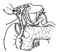
A schematic drawing of the extent of lymphadenectoA schematic drawing of the extent of lymphadenectomy for gallbladder cancer, especially when the extrahepatic biliary tree is resected.

Benign lesions

Benign lesions of the gallbladder are relatively common, but only adenomatous polyps are considered to have malignant potential. Although ultrasonography can be useful in evaluating these lesions, considerable difficulty may be encountered in establishing the diagnosis preoperatively.

Cholesterol polyps

Cholesterol polyps account for approximately 50% of all polypoid lesions of the gallbladder.
These lesions are thought to originate from a defect in cholesterol metabolism. They appear as yellow spots on the mucosal surface of the gallbladder and are identified histologically as epithelial-covered macrophages laden with triglycerides and esterified sterols in the lamina propria of the mucosal layer of the gallbladder.
As a rule, cholesterol polyps exist as multiple lesions and are usually less than 10 mm. Cholesterol polyps are generally asymptomatic.

Inflammatory polyps

These lesions result from chronic inflammation. They extend into the gallbladder lumen by a narrow vascularized stalk.

Adenomyomatosis

Adenomyomatosis is characterized by extensions of Rokitansky-Aschoff sinuses through the muscular wall of the gallbladder. Ultrasonography reveals a thickened gallbladder wall with intramural diverticula. Although adenomyomatosis is generally considered a benign condition, serial evaluation with ultrasonography is indicated to rule out enlarging adenomatous polyps and gallbladder cancer. Some authors have reported gallbladder cancer occurring in localized adenomyomatosis and have suggested a more aggressive approach to the benign lesions.

Adenomatous polyps

Adenomatous polyps are benign epithelial neoplasms with malignant potential. Papillary adenomas grow as pedunculated, complex, branching tumors projecting into the gallbladder lumen. Tubular adenomas arise as a flat, sessile neoplasm. Consequently, it can be difficult to distinguish some adenomas from other gallbladder polyps by ultrasonography. Like many gastrointestinal tumors, an adenoma-carcinoma sequence is generally thought to occur in these lesions.

Other lesions

Other rare, benign lesions found in the gallbladder include fibromas, leiomyomas, lipomas, hemangiomata, granular cell tumors, and heterotropic tissue, including gastric, pancreatic, and intestinal epithelium.

Malignant lesions

The incidence of gallbladder cancer is 1.2 cases 100,000 persons in the United States; the frequency is much higher in Mexican Americans and Native Americans, although the greatest incidence is found in the indigenous peoples of the Andes Mountains, in northeastern Europeans, and in Israelis. The female-to-male ratio for gallbladder cancer is about 3:1; incidence of the disease peaks in the seventh decade of life.[1]
The most common risk factor for gallbladder cancer is gallstones, which are present in 75%-90% of gallbladder cancer cases. The size of the gallstones plays a role in the risk of developing of gallbladder cancer. Gallbladders containing gallstones that are greater than 3 cm in diameter have a 10-fold greater risk for developing malignancy than do those containing gallstones that are 1 cm in diameter. Causality is difficult to establish, but other chronic inflammatory conditions, such as cholecystoenteric fistula, primary sclerosing cholangitis, pancreaticobiliary maljunction, and chronic infection with Salmonella typhi, have also been associated with an increased risk of gallbladder cancer.
Modern series report about a 10% incidence of gallbladder cancer in porcelain gallbladders (in which the gallbladder wall is calcified), a much lower rate than that reported in older series. Stippled calcification of the mucosa is thought to carry a higher risk of gallbladder cancer than does generalized calcification of the gallbladder wall.[2, 3] Based on these associations, chronic inflammation is postulated to be involved in the pathogenesis of gallbladder cancer.
Gallbladder cancer is often discovered incidentally during a workup for gallstone disease, and about 50% of gallbladder cancer cases are diagnosed incidentally in cholecystectomy specimens. Unfortunately, about 35% of patients have distant metastases at the time of diagnosis.
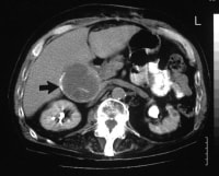
Histologically, adenocarcinoma is found in 90% of gallbladder cancer cases, and squamous cell carcinoma is found in 2% of cases. Rare types of gallbladder cancer include sarcoma, adenosquamous carcinoma, oat cell carcinoma, carcinoid, lymphoma, melanoma, and metastatic tumors. A number of histologic subtypes of adenocarcinoma have been described, but papillary adenocarcinoma represents about 5% of gallbladder cancers; it tends to be well-differentiated and carries a more favorable prognosis. (See images below.)
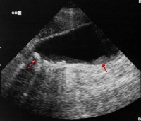
Sagittal ultrasonogram in a 71-year-old woman. ThiSagittal ultrasonogram in a 71-year-old woman. This image demonstrates heterogeneous thickening of the gallbladder wall (arrows). The diagnosis was primary papillary adenocarcinoma of the gallbladder. A transaxial enhanced computed tomography (CT) scaA transaxial enhanced computed tomography (CT) scan of a 60-year-old man with right upper quadrant pain shows a partially calcified gallbladder (arrow). At laparotomy and histology, an infiltrating adenocarcinoma of the gallbladder was confirmed.
Gastroenterology


Biliary Disease

A diverse spectrum of disease affects the biliary system, often presenting with similar clinical signs and symptoms.
A normal postcholecystectomy cholangiogram. A normal postcholecystectomy cholangiogram. Biliary disease. In this patient with persistent eBiliary disease. In this patient with persistent elevation of liver-associated enzymes, the contrast entering the biliary ductal system preferentially enters the cystic duct. Biliary disease. Even when the catheter is advanceBiliary disease. Even when the catheter is advanced to the proximal common hepatic duct, contrast dye preferentially fills the cystic duct and gallbladder rather than allowing visualization of the intrahepatic ductal system. Biliary disease. In this image, the common bile duBiliary disease. In this image, the common bile duct is occluded with a balloon-tipped catheter. Contrast fills the intrahepatic ductal system to reveal diffuse intrahepatic sclerosing cholangitis.

Dermatitis

Background

Berloque dermatitis obtains its name from the German word berlock or the French berloque, meaning trinket or charm. Rosenthal[1] coined the term in 1925 to describe pendantlike streaks of pigmentation on the neck, face, arms, or trunk. He suspected they were due to fluid droplets, unaware that Freund[2] in 1916 had described hyperpigmented macules due to sun exposure after the application of eau de cologne. The phototoxic ingredient causing the pigmentation proved to be bergapten, a component of oil of bergamot, derived from the rind of Citrus bergamia, the bergamot lime. Several cases were reported in the 1950s and 1960s following increased use of perfumes containing oil of bergamot and the passion for sunbathing. Since the introduction of artificial oil of bergamot and the reduced use of the natural product in perfumes, berloque dermatitis has become rare. Note the image below.
Hyperpigmented streaks on the dorsa of hands of a Hyperpigmented streaks on the dorsa of hands of a patient with bergapten phototoxicity Related eMedicine articles include Cosmetics and Cosmeceuticals.

Allergy and Immunology

Allergic Fungal Sinusitis

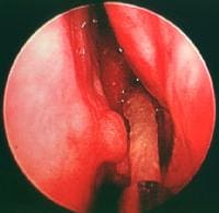
Over the past 2 decades, allergic fungal sinusitis (AFS) has become increasingly defined. Historically mistaken for a paranasal sinus tumor, allergic fungal sinusitis (AFS) now is believed to be an allergic reaction to aerosolized environmental fungi, usually of the dematiaceous species, in an immunocompetent host. This is in contrast to invasive fungal infections that affect immunocompromised hosts, such as patients with diabetes mellitus and patients with AIDS. Most patients with allergic fungal sinusitis (AFS) have a history of allergic rhinitis, and the exact timing of allergic fungal sinusitis (AFS) development can be difficult to discern. Thick fungal debris and mucin, as shown below, are developed in the sinus cavities and must be surgically removed so that the inciting allergen is no longer present. Recurrence is not uncommon once the disease is removed. Anti-inflammatory medical therapy and immunotherapy are being employed to help prevent recurrence.
Left middle meatus with suctioning of thick allergLeft middle meatus with suctioning of thick allergic mucin from the ethmoid bulla in the center of the picture; the end of the suction is in the inferior portion of the picture.