الجمعة، 3 فبراير 2012

Bile Duct Tumors

Tumors of the biliary tract (see image below) are uncommon but serious problems. The spectrum of lesions ranges from benign tumors, such as adenomas, to malignant lesions, such as adenocarcinomas. This discussion excludes tumors of the gallbladder, which are discussed separately.
Distal common bile duct tumor excised by radical pDistal common bile duct tumor excised by radical pancreaticoduodenectomy. The tumor measured 1.2 cm in diameter.
Most patients with bile duct tumors present with jaundice due to obstruction of the biliary tree by the tumor. Because the tumors are generally small, standard imaging studies, such as ultrasonography[1] and computed tomography (CT) scanning, may fail to show the lesion. These techniques may, however, provide a clue to the level of the obstruction and help exclude metastatic disease.
Cholangiography via a transhepatic or endoscopic approach is required to define the biliary anatomy and extent of the lesion. Magnetic resonance cholangiography is a noninvasive alternative available in an increasing number of centers.
The anticipated course of most cases of bile duct tumors includes recurrent biliary obstruction with infectious complications, local spread, and death in 6-12 months. Treatment depends on the site and extent of the lesion, and surgical resection improves survival and prognosis.

Benign Gastric Tumors

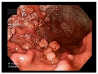
With the advent of modern techniques and the widespread use of gastric endoscopy, benign gastric wall lesions are now diagnosed more frequently and can be studied using the tissue obtained by biopsy or polypectomy. In the past, the diagnosis of gastric tumors was based on x-ray examination, but, in 1922, Schendler was the first to make an endoscopic-based diagnosis.[1] Note the image below.
Upper endoscopy showing multiple gastric polyps. Upper endoscopy showing multiple gastric polyps.

Acute Mesenteric Ischemia

Acute mesenteric ischemia (AMI) is a syndrome in which inadequate blood flow through the mesenteric circulation causes ischemia and eventual gangrene of the bowel wall. Broadly, AMI may be classified either as arterial or venous disease.
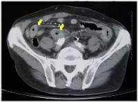
Arterial disease may be subdivided into nonocclusive mesenteric ischemia (NOMI; see the image below) and occlusive mesenteric arterial ischemia (OMAI). OMAI may be further subdivided into acute mesenteric arterial embolus (AMAE) and acute mesenteric arterial thrombosis (AMAT). Venous disease takes the form of mesenteric venous thrombosis (MVT). Thus, for practical purposes, AMI comprises 4 different primary clinical entities: NOMI, AMAE, AMAT, and MVT.
CT scan (with contrast) of nonocclusive mesentericCT scan (with contrast) of nonocclusive mesenteric ischemia with resulting bowel wall edema (arrows). The 4 types of AMI have somewhat different predisposing factors, clinical pictures, and prognoses. A secondary clinical entity of mesenteric ischemia occurs because of mechanical obstruction, such as internal hernia with strangulation, volvulus, intussusception, tumor compression, and aortic dissection. Occasionally, blunt trauma may cause isolated dissection of the superior mesenteric artery (SMA) and lead to intestinal infarction.
Because the 4 types of AMI share many similarities and a final common pathway (ie, bowel infarction and death, if not properly treated), they are discussed together.
In 1930, Cokkinis remarked, “Occlusion of the mesenteric vessels is apt to be regarded as one of those conditions of which the diagnosis is impossible, the prognosis hopeless, and the treatment almost useless.”[1] This quote indicates some of the extreme difficulties faced by physicians treating AMI. Symptoms are nonspecific initially, before evidence of peritonitis presents. Thus, diagnosis and treatment are often delayed until the disease is advanced.
Fortunately, since 1930, many advances have been made that allow earlier diagnosis and treatment. Whereas the prognosis remains grave for patients in whom the diagnosis is delayed until bowel infarction has already occurred, patients who receive the appropriate treatment in a timely manner are much more likely to recover.[2]

Abdominal Angina

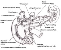
Abdominal angina is defined as the postprandial pain that occurs in individuals with sufficient mesenteric vascular occlusive disease such that blood flow cannot increase enough to meet visceral demands. The mechanism is believed to be similar to the angina pectoris that occurs in individuals with coronary artery disease or the intermittent claudication that accompanies peripheral vascular disease, as depicted in the image below.
The superior mesenteric artery and inferior mesentThe superior mesenteric artery and inferior mesenteric artery share collateral circulation near the splenic flexure of the colon. When dilated, this vessel is termed the meandering mesenteric artery. As seen on an angiogram, this is a sign of chronic mesenteric ischemia. The pancreaticoduodenal arcades are collateral patThe pancreaticoduodenal arcades are collateral pathways between the celiac artery and the superior mesenteric artery.
Although Schnitzler first described the clinical picture of postprandial clinical pain in 1901, description of the syndrome of postprandial abdominal angina generally is attributed to Baccelli or Goodman (1918). In 1936, Dunphy recognized that this syndrome was a precursor of fatal intestinal necrosis; however, not until 1957 did Mikkelsen propose surgical treatment of occlusive mesenteric vascular disease. Shaw and Maynard reported the first transarterial thromboendarterectomy of the superior mesenteric artery (SMA) in 1958, followed in rapid succession by Mikkelsen and Zarro in 1959. Numerous technical refinements followed.

Lower Gastrointestinal Bleeding

Lower gastrointestinal bleeding (LGIB) accounts for approximately 20-33% of episodes of gastrointestinal (GI) hemorrhage, with an annual incidence of about 20-27 cases per 100,000 population in Western countries. However, although LGIB is statistically less common than upper GI bleeding (UGIB), it has been suggested that LGIB is underreported because a higher percentage of affected patients do not seek medical attention.[1] Indeed, LGIB continues to be a frequent cause of hospital admission and is a factor in hospital morbidity and mortality LGIB is distinct from UGIB in epidemiology, management, and prognosis.
LGIB encompasses a wide spectrum of symptoms, ranging from trivial hematochezia to massive hemorrhage with shock. Acute LGIB is defined as bleeding that is of recent duration, originates beyond the ligament of Treitz, results in instability of vital signs, and is associated with signs of anemia with or without need for blood transfusion.
LGIB is classified under 3 groups according to the amount of bleeding, as shown in the image below. Massive hemorrhage is a life-threatening condition and requires transfusion of at least 5 U of blood.
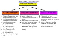
Types of lower gastrointestinal (GI) bleeding. HR Types of lower gastrointestinal (GI) bleeding. HR = heart rate; SBP = systolic blood pressure.
Massive lower GI bleeding is defined as follows:
  • Passage of a large volume of red or maroon blood through the rectum
  • Hemodynamic instability and shock
  • Initial decrease in hematocrit (Hct) level of 6 g/dL or less
  • Transfusion of at least 2 U of packed red blood cells (RBCs)
  • Bleeding that continues for 3 days
  • Significant rebleeding in 1 week
LGIB has a mortality rate ranging from about 10-20%, with patients of advanced age (>60 y) and patients with comorbid conditions (eg, multiorgan system disease, transfusion requirements in excess of 5 units [U], need for operation, and recent stress, such as surgery, trauma, and sepsis) at greatest risk. LGIB is more likely in the elderly because of a higher incidence of diverticulosis and vascular disease in these groups. The incidence of LGIB is higher in men than in women.
Advances in diagnostic and therapeutic colonoscopy and in interventional angiography have resulted in a shift away from the need for surgical treatment (see the image below). Effective management with less invasive modalities has also reduced healthcare costs and, more importantly, patient morbidity and mortality.
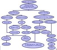
Methods used to treat lower gastrointestinal bleedMethods used to treat lower gastrointestinal bleeding (LGIB).

Historical details

Understanding of the pathogenesis, diagnosis, and treatment of LGIB has drastically changed during the last 50 years. In the first half of the 20th century, large intestinal neoplasms were believed to be the most common cause of LGIB. In the 1950s, this condition was commonly attributed to diverticulosis; surgical treatment consisted of blind segmental bowel resections, with disappointing results. Patients who underwent these procedures suffered from a prohibitively high rebleeding rate (up to 75%), morbidity (up to 83%), and mortality (up to 60%).
In the last 4 decades, diagnostic methods for locating the precise bleeding point greatly improved. The flexible endoscope was developed in 1954. The full-length colonoscope was developed in 1965 in Japan. Also in 1965, Baum et al described selective mesenteric angiography, which permitted the identification of vascular abnormalities and the precise bleeding point.[2] The first anal colonoscopy was performed in 1969.
Experience with mesenteric angiography in the late 1960s and 1970s suggested that angiodysplasias and diverticulosis were the most common reasons for LGIB. Since its discovery, mesenteric angiography remains the criterion standard in precise localization of the bleeding.
Rosch et al described superselective visceral arteriography for infusion of vasoconstrictors in 1971 and superselective embolization of the mesenteric vessels as an alternative technique to treat massive LGIB in 1972.[3, 4] The most feared complication of embolization of the mesenteric vessels is ischemic colitis, which has limited its use for GI bleeding.
The initial experience with vasopressin infusion was reported in 1973-1974. Vasopressin causes vasoconstriction and arrests the bleeding in 36-100% of patients. The recurrence rate following completion of vasopressin infusion can be as high as 71%; therefore, vasopressin is used to temporize the acute event and to stabilize patients before surgery.
Endoscopic control of bleeding with thermal modalities or sclerosing agents has been in use since the 1980s. One of the advantages of upper (or lower) endoscopic evaluation is that it provides a means to administer therapy in patients with GI bleeding. Nuclear scintigraphy has been used since the early 1980s as a very sensitive diagnostic tool to evaluate bleeding from GI tract; this modality can detect hemorrhage at rates as low as 0.1 mL/min
Familial Adenomatous Polyposis

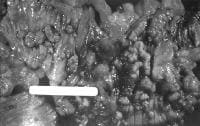
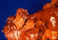
Familial adenomatous polyposis (FAP) is the most common adenomatous polyposis syndrome. It is an autosomal dominant inherited disorder characterized by the early onset of hundreds to thousands of adenomatous polyps throughout the colon. If left untreated, all patients with this syndrome develop colon cancer by age 35-40 years. In addition, an increased risk exists for the development of other malignancies. See the image below.
Colectomy specimen obtained from a patient with faColectomy specimen obtained from a patient with familial adenomatous polyposis. Note the presence of numerous synchronous adenomatous polyps lining the luminal surface. The genetic defect in FAP is a germline mutation in the adenomatous polyposis coli (APC) gene. Syndromes once thought to be distinct from FAP are now recognized to be, in reality, part of the phenotypic spectrum of FAP.
Syndromes with a germline mutation in the APC gene include FAP, Gardner syndrome, some families with Turcot syndrome, and attenuated adenomatous polyposis coli (AAPC). Gardner syndrome is characterized by colonic polyposis typical of FAP, along with osteomas (bony growth most commonly on the skull and the mandible), dental abnormalities, and soft tissue tumors. Turcot syndrome is characterized by colonic polyposis typical of FAP, along with central nervous system tumors (medulloblastoma). AAPC is characterized by fewer colonic polyps (average number of polyps, 30-35) as compared to classic FAP. The polyps also tend to develop at a later age (average age, 36 y), and they tend to involve the proximal colonic area.
In considering the spectrum of polyposis syndromes, patients with multiple adenomatous polyps most likely have FAP (or one of its variants), AAPC, or MYH-associated polyposis (MAP). If a patient with a suspected polyposis syndrome undergoes genetic testing and does not have an APC gene mutation, MYH gene testing should be performed to assess for MAP, as 10-20% of patients who do not have an APC gene mutation have biallelic MYH gene mutations.[1]
The phenotype of MAP is often indistinguishable from FAP or AAPC, with patients having usually 10-100 polyps but sometimes more than 100. The age of onset of MAP is usually in patients older than 45 years, and patients often present symptomatically, with colorectal carcinoma commonly found at the time of diagnosis. This is in part because there is usually no family history given the autosomal recessive inheritance pattern of MAP. Duodenal polyps can be found in up to one fifth of patients.[2] There is no increased risk of other types of cancers associated with this syndrome.

Schatzki Ring

Since the 1950s, several investigators have published reports of patients with dysphagia who had associated lower esophageal ringlike constrictions, but each investigator had a different opinion as to the cause and nature of these rings. In 1953, Ingelfinger and Kramer believed that these rings occurred as a result of a contraction by an overactive band of esophageal muscle[1] ; however, Schatzki and Gary believed that these rings were fixed and not contractile.[2] Some of this controversy may be related to the confusion of categorizing muscular and mucosal rings under the same entity, as concluded by Goyal et al.[3, 4, 5, 6]
Endoscopic appearance of the distal esophagus illuEndoscopic appearance of the distal esophagus illustrating a Schatzki ring.
Two rings have been identified in the distal esophagus. The muscular ring, or A ring, is a thickened symmetric band of muscle that forms the upper border of the esophageal vestibule and is located approximately 2 cm above the gastroesophageal junction. The A ring is rare; furthermore, it is even more rarely associated with dysphagia. On the other hand, the mucosal ring, or B ring, is quite common and is the subject of discussion in this article. The B ring is a diaphragmlike thin mucosal ring usually located at the squamocolumnar junction; it may be symptomatic or asymptomatic, depending on the luminal diameter.
The pathogenesis is not clear, and patients typically present with intermittent nonprogressive dysphagia for solids. Fortunately, most patients respond well to initial and repeat dilatation therapy. A small number of patients may have stubborn rings that require more aggressive endoscopic or surgical intervention.